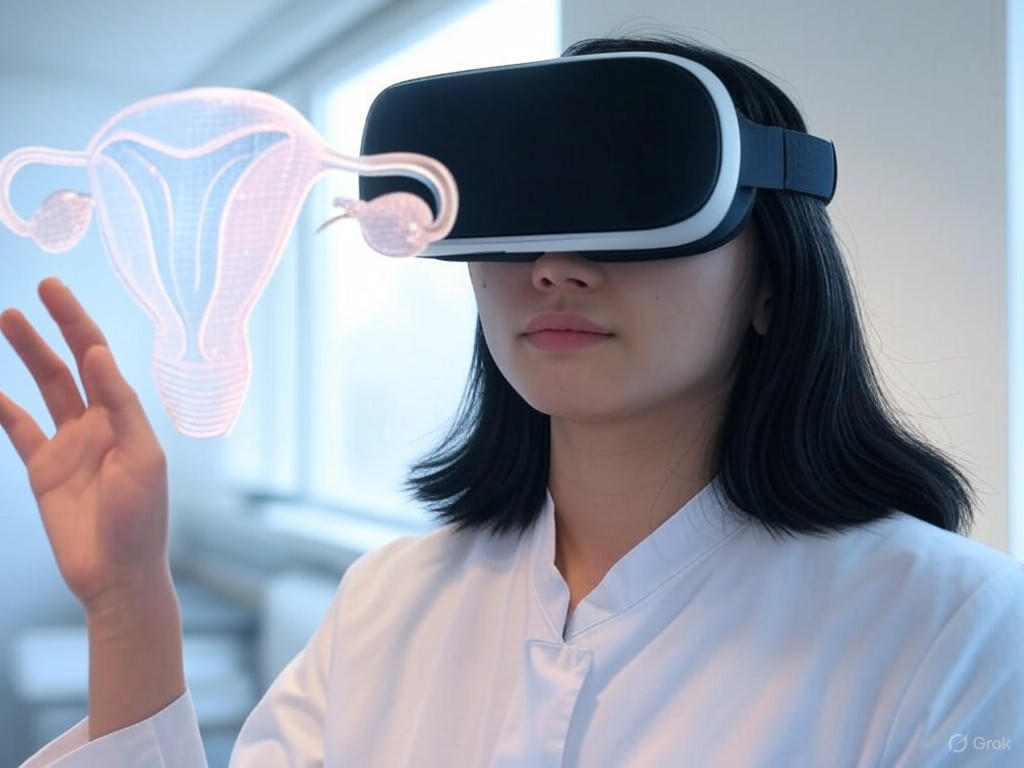
Experiencing or preparing for medical procedures involving the uterus can feel overwhelming, especially when the anatomy feels abstract or difficult to visualize. Many individuals struggle to fully understand their reproductive health due to limited access to clear, interactive tools that demystify complex anatomical structures. This lack of clarity can lead to anxiety or uncertainty when facing medical decisions or exploring options like fertility treatments or gynecological care.
3D uterus models in virtual reality (VR) offer a groundbreaking way to visualize and explore your body’s inner workings. This technology is transforming how patients, partners, and medical professionals approach reproductive health education and treatment. This article explores how 3D uterus models in VR empower you to visualize your anatomy in an immersive, accessible way. We’ll dive into how this technology works, its benefits for education and medical consultations, and how it’s helping bridge the gap between patients and their reproductive health.
- Why are 3D VR uterus models redefining fertility education?
- How are medical scans turned into interactive 3D uterus models?
- Which VR platforms let you explore the uterus—and what do they cost?
- How can VR uterus walkthroughs support patients during fertility treatment?
- What do students and doctors gain from VR uterus simulations in training?
- Are personalized uterus models a reality yet?
- What equipment and setup do you need to view your uterus in VR?
- What limitations and future breakthroughs should users know about?
- Limitations:
- Future Breakthroughs:
- Your top questions, answered
- Final thoughts
- References
Why are 3D VR uterus models redefining fertility education?
3D virtual reality (VR) uterus models are transforming fertility education by making complex anatomical and reproductive concepts more accessible and engaging. These immersive tools allow users to interactively explore the uterus and related structures, which enhances understanding, knowledge retention, and self-efficacy compared to traditional teaching methods. Studies show that VR models help patients and students visualize intricate processes, such as embryological development and pelvic floor disorders, at their own pace and on various devices, leading to improved comprehension and motivation for positive behavioral change.
In medical training, 3D-printed uterus models combined with VR and realistic silicone components provide hands-on practice for gynecologic procedures, improving technical skills, communication, and patient-centered care. The affordability and replicability of these models further support their widespread adoption in educational settings. Overall, 3D VR uterus models bridge gaps in understanding, empower learners and patients, and hold promise for better outcomes in fertility and women’s health education.
If you’d like a taste of this tech in action, our VR IVF Lab Tour shows how one clinic weaves virtual walkthroughs into each visit.

How are medical scans turned into interactive 3D uterus models?
Turning your medical scans into interactive 3D uterus models is a step-by-step process that brings your anatomy to life. Here’s how it works:
- First, you get a scan; usually Magnetic Resonance Imaging (MRI) or Computed Tomography (CT). These scans capture detailed images of your uterus in thin slices.
- Next, experts use special software to “segment” the uterus. This means they carefully outline the uterus and any other important structures, separating them from the rest of the image.
- The software then stacks these slices and creates a digital 3D model. This model can be refined for accuracy and smoothed out for a realistic look.
- Now, your 3D uterus can be explored on a screen, in virtual reality, or even printed as a physical model. You can zoom, rotate, and interact with it; making it easier to understand your own body.
- This technology helps you and your care team visualize conditions, plan treatments, and communicate clearly. As Dr. Zhonghua Sun, PhD, Professor of Medical Imaging, says: “3D models based on your own scans make complex anatomy easier to understand and support shared decision-making in your care”.
The American College of Obstetricians and Gynecologists (ACOG) encourages using clear, patient-centered tools like these to empower you in your pregnancy journey. Interactive 3D models can help you feel more confident and informed about your health.
Which VR platforms let you explore the uterus—and what do they cost?
Several VR platforms offer uterus exploration experiences. Here’s a simple table to help you compare VR (virtual reality) platforms for exploring the uterus, including what you’ll need and the typical cost:
| Platform/Tool | What You Need | Typical Cost | Notes |
|---|---|---|---|
| Smartphone VR (e.g., Google Cardboard) | Your smartphone + cardboard viewer | $10–$30 | Affordable, easy to use, use free/low-cost apps 1. |
| Advanced VR Headsets (e.g., Oculus Rift, Meta Quest) | VR headset + compatible computer or standalone | $300–$500+ | Immersive, used in clinics/universities, costly 3. |
| Custom Medical Platforms (e.g., Elucis) | Specialized VR system, often in clinics | Varies (clinic-based) | Used for detailed medical imaging 5 |
Dr. Christian Moro, PhD, says, “the more cost-effective mobile-based VR was just as suitable for teaching isolated systems as the more expensive desktop-based VR”.
Most people start with smartphone VR for convenience and price. If you want a deeper experience, advanced headsets are available in some clinics or for home use. Always check with your care team about what’s available and best for you.
Some premium apps now sync menstrual-cycle visuals with calendar data pulled from your favorite tracker—many of those top choices appear in our Top Fertility Apps 2025 guide.
How can VR uterus walkthroughs support patients during fertility treatment?
Virtual reality (VR) uterus walkthroughs can be a powerful support tool during your fertility treatment. They help you see and understand what’s happening inside your body, making complex steps feel less mysterious and more manageable. Here’s how they can help you:
-
You get a clear, interactive view of your uterus and reproductive system, so you can better understand procedures and treatment plans.
-
VR walkthroughs can lower your anxiety and help you feel more in control, especially during stressful moments in your fertility journey.
-
Many people find that VR makes it easier to ask questions and talk with their care team, leading to more confident, informed decisions.
-
For some, VR experiences offer a calming distraction during procedures, with 90% of participants in one study saying it helped reduce their anxiety and 71% reporting it distracted them from pain .
-
VR can be tailored to your needs, including mindfulness and relaxation exercises, which support your mental health and resilience.
Dr. Anna Alexandra McDougall, MBBS, who led a recent study, says, “VR is a feasible and acceptable option that may improve patient experience during uterine procedures—even if it doesn’t always lower pain scores, most people feel less anxious and more supported”.
What do students and doctors gain from VR uterus simulations in training?
When students and doctors use virtual reality (VR) uterus simulations in training, they gain practical skills and confidence that help them care for you more safely and compassionately. Here’s what these simulations offer:
- You get doctors and nurses who have practiced procedures in a realistic, risk-free environment, so they’re better prepared for real-life situations.
- Students learn to recognize and manage emergencies, like cesarean sections, with higher confidence and better test scores. One study showed VR-trained participants scored 17% higher on knowledge tests than those using traditional methods.
- VR lets learners repeat scenarios as often as needed, building muscle memory and reducing anxiety when facing similar situations with real patients.
- These simulations encourage teamwork and communication, which are key for safe, patient-centered care.
- VR training is highly accepted by students and doctors, who report feeling more motivated and engaged.
As Dr. Hyeon Ji Kim, MD, notes, “VR simulation can be an effective educational tool for improving participants’ knowledge and confidence in managing patients and performing procedures”.
The American College of Obstetricians and Gynecologists (ACOG) supports using advanced, hands-on training tools like VR to ensure your care team is ready for anything. This means you benefit from providers who are skilled, confident, and up-to-date with the latest technology.
Are personalized uterus models a reality yet?
Yes, personalized uterus models are now a reality—and they’re making a real difference in care and understanding. Using your own medical scans, such as three-dimensional ultrasound or magnetic resonance imaging (MRI), specialists can create a detailed, 3D-printed model of your uterus. These models are already being used for:
- Patient education and counseling, so you can see and touch a model that matches your own anatomy.
- Surgical planning, especially for complex cases like uterine anomalies or multiple fibroids, helping your care team prepare and explain procedures to you.
- Medical training, giving students and doctors hands-on practice with realistic, patient-specific models.
- A recent 2024 study confirms, “The creation of personalized 3D-printed uterine models for utilization in reproductive endocrinology and infertility is feasible and offers a valuable tool for both patients and providers”.
These models are accurate—errors are typically less than 2 millimeters—and patients report they help with understanding and decision-making.
What equipment and setup do you need to view your uterus in VR?
To view your uterus in virtual reality (VR), you’ll need a few key pieces of equipment and a simple setup. Here’s what you should have:
- A VR headset: This can be a basic, affordable option like Google Cardboard (used with your smartphone), or a more advanced headset such as Meta Quest or Oculus Rift. Prices range from $10 for cardboard viewers to $300+ for advanced headsets.
- A compatible device: For basic VR, your smartphone is enough. For advanced headsets, you may need a computer or a standalone VR device.
- Specialized VR app or program: Look for apps designed for reproductive health or uterus visualization. Some clinics may offer custom programs that use your own medical images.
- Optional: Medical imaging data: For a truly personalized experience, your care team might use your ultrasound or magnetic resonance imaging (MRI) scans to create a 3D model of your uterus.
Setup is usually simple: download the app, place your phone in the viewer or connect your headset, and follow the on-screen instructions. Some clinics use more advanced systems that sync VR with real-time uterine activity for pain management and education.
Dr. Antonio Melillo, MD, explains, “VR may be considered as an effective nonpharmacological technique for the treatment of pain and anxiety and fear of childbirth experience during labor”. Always ask your provider which options are available and best for you.
What limitations and future breakthroughs should users know about?
Limitations:
When using virtual reality (VR) to view your uterus, there are some important challenges to keep in mind.
- Not everyone feels comfortable using VR. In one study, only about two-thirds of people who were offered a VR video actually chose to watch it.
- VR may not lower anxiety for everyone. The overall reduction in anxiety before a clinic visit was not significant compared to standard information.
- People who already feel more anxious may be less likely to use VR tools.
- Some users may find the technology unfamiliar or even uncomfortable.
Future Breakthroughs:
Looking ahead, VR has the potential to become a more helpful and inclusive tool for reproductive health.
- More personalized and interactive VR experiences could help more people feel comfortable and engaged.
- Improved education tools may better support those with higher anxiety.
- As VR becomes more common, clinics may offer more tailored content, making it easier for you to understand your care and feel empowered.
- Ongoing research is exploring how VR can be used for pain management, birth preparation, and even real-time support during labor 1.
Your top questions, answered
What exactly is a 3D uterus model in VR?
A 3D uterus model in virtual reality (VR) is a digital, interactive representation of the uterus that you can explore using VR headsets or screens. These models are created from medical imaging or bioprinting data and can show the uterus’s layers, blood vessels, and even conditions like endometriosis. They help you and your care team visualize anatomy for education, planning, or support.
Do these models improve fertility outcomes?
3D models and bio-printed constructs are showing promise for repairing damaged uterine tissue and improving fertility in research settings. For example, a 3D bio-printed endometrial construct restored fertility in 75% of rats with severe uterine injury, compared to just 12.5% with standard treatment. However, using 3D ultrasound to measure the uterus hasn’t been shown to predict pregnancy success in in vitro fertilization (IVF) cycles. More research is needed before these models can guarantee better outcomes for everyone.
Where can I download a free uterus model?
Free 3D uterus models are available on open-source platforms like NIH 3D Print Exchange or Sketchfab. These models are for educational use and may not reflect your unique anatomy. Always check with your healthcare provider before using any model for medical decisions.
Can I visualize conditions like endometriosis in VR?
Yes, VR and 3D imaging can help visualize conditions like endometriosis by showing the uterus’s structure and muscle layers in detail. This can help you understand your diagnosis and treatment options, but these tools are still being developed and may not capture every detail of your condition.
Is VR safe during pregnancy?
Current research suggests that using VR for viewing 3D models or for education is safe during pregnancy, as it does not involve radiation or invasive procedures. If you experience dizziness or discomfort, take breaks and talk to your provider.
Final thoughts
Virtual reality (VR) is opening new doors for how you connect with your pregnancy and manage your care. VR can help you see your baby in new ways, ease pain and anxiety during labor, and make medical information more understandable and personal. While not everyone finds VR helpful for anxiety or pain, most people who try it say it adds value to their experience.
If you’re curious, ask your provider about VR options—they’re safe to use during pregnancy and may help you feel more connected and informed. As technology improves, you’ll see even more ways VR can support you, from visualizing your baby’s growth to managing labor pain in real time. Your comfort and empowerment matter, and VR is one more tool to help you on your journey.
References
-
The effect of an informative 360-degree virtual reality video on anxiety for women visiting the one-stop clinic for abnormal uterine bleeding: A randomized controlled trial (VISION-trial)… European journal of obstetrics, gynecology, and reproductive biology. 2022; 272. https://doi.org/10.1016/j.ejogrb.2022.02.179
-
Synchronization of a Virtual Reality Scenario to Uterine Contractions for Labor Pain Management: Development Study and Randomized Controlled Trial… Games for health journal. 2024 https://doi.org/10.1089/g4h.2023.0202
-
Personalized medicine in the evaluation of Müllerian anomalies: the role of three-dimensional printing technology. F&S Reports. 2024; 5. https://doi.org/10.1016/j.xfre.2024.05.003
-
Immersive virtual reality simulation training for cesarean section: a randomized controlled trial. International Journal of Surgery (London, England), 110, 194 - 201. https://doi.org/10.1097/JS9.0000000000000843
-
Virtual reality for the management of pain and anxiety during outpatient manual vacuum aspiration for miscarriage or incomplete abortion: a mixed methods trial. The European Journal of Contraception & Reproductive Health Care, 29, 298 - 304. https://doi.org/10.1080/13625187.2024.2410838
-
Patient-Specific 3D-Printed Low-Cost Models in Medical Education and Clinical Practice. Micromachines, 14. https://doi.org/10.3390/mi14020464.
-
Fetal heart segmentation in a virtual reality environment… The international journal of cardiovascular imaging. https://doi.org/10.1007/s10554-024-03157-0.
-
Design of virtual reality for teaching ultrasound in medical and engineering education. The Journal of the Acoustical Society of America. https://doi.org/10.1121/10.0007617.
-
An affordable platform for virtual reality-based patient education in radiation therapy. Practical radiation oncology. https://doi.org/10.1016/j.prro.2023.06.008.